Gallery

Understanding Hallux Valgus Through Visuals: Medical X-rays & More

Explore our comprehensive image gallery dedicated to Hallux valgus—commonly known as bunions. Visual learning can make all the difference when it comes to understanding complex foot deformities. That’s why we’ve gathered a wide range of real medical images, including X-rays, clinical photos, and detailed diagrams, to help you see exactly what Hallux valgus is and how it progresses.

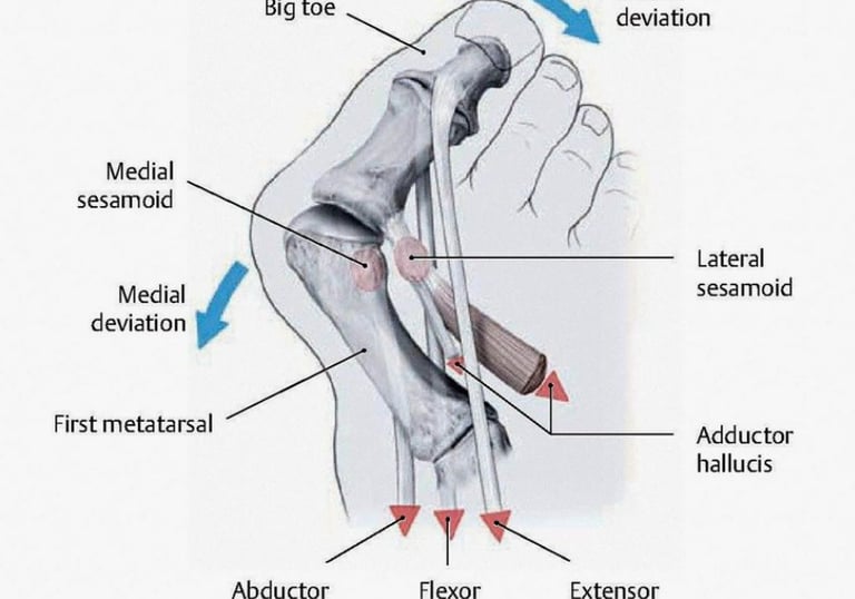
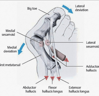
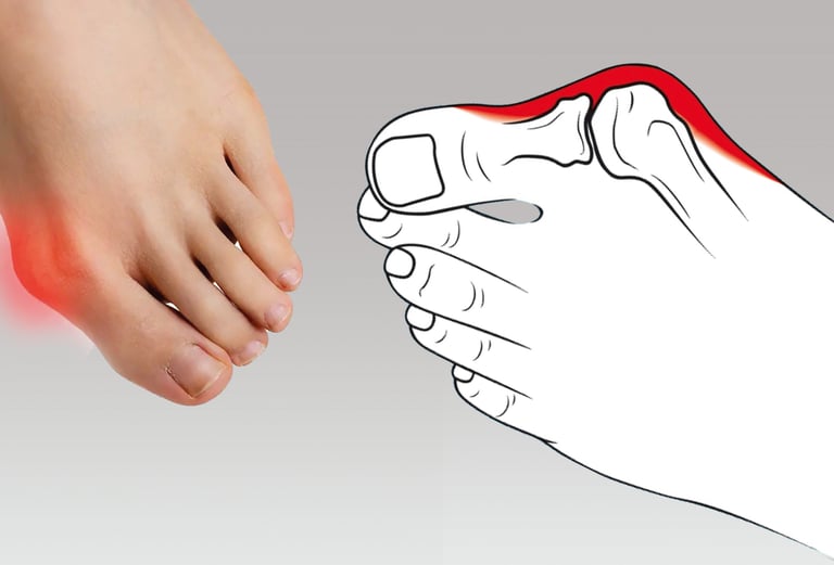
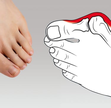
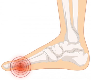
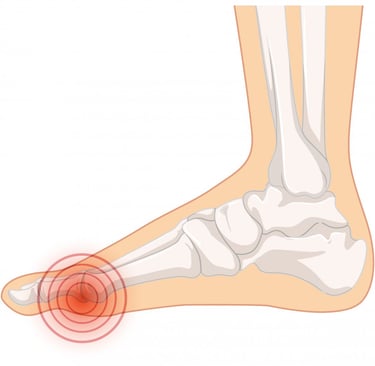
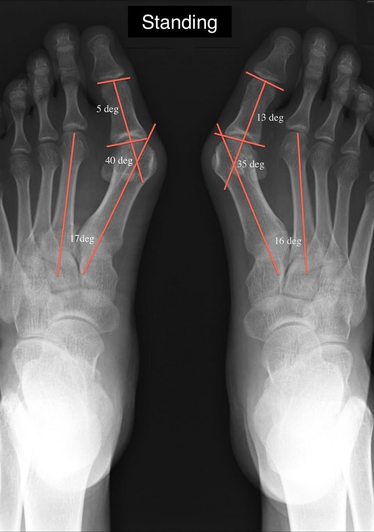
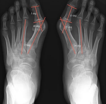
X-ray images provide a unique window into the underlying bone structure of the foot. By viewing these, you can easily recognize the characteristic outward shift of the big toe (hallux) and the development of the bunion at the joint. Clinical photographs showcase how the deformity looks externally, while annotated diagrams explain key anatomical features and the changes that occur over time.

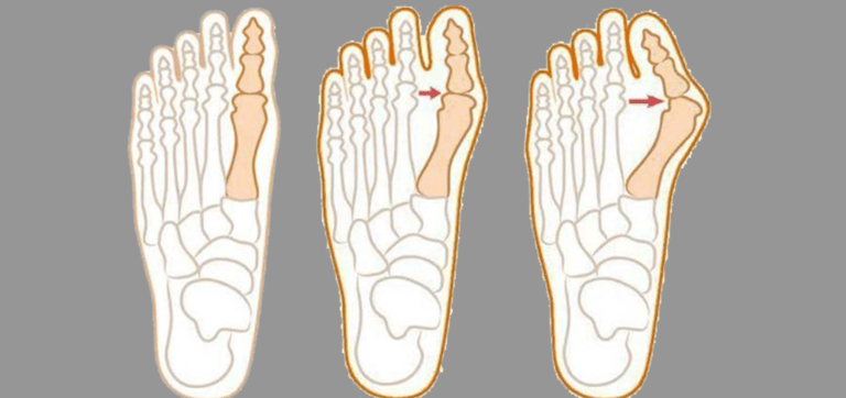
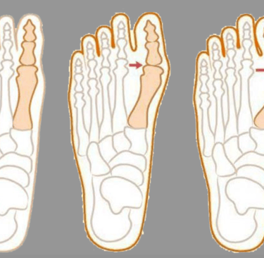
Whether you’re a patient, a student, or just curious, our gallery allows you to compare different stages of bunion development, from mild to severe. You’ll see pre- and post-operative images, examples of conservative treatment devices (like toe spacers and orthotic splints), and variations in bunion appearance across different individuals.

Visual resources like these help you make more informed decisions about your own care or that of a loved one. They also make conversations with your healthcare provider clearer, since you can point out exactly what you notice in your own foot.

Use this gallery to support your understanding of Hallux valgus and explore the possibilities for management and treatment. Visual learning isn’t just helpful—it’s essential for making sense of medical conditions like bunions.

A detailed anatomical model of the human foot showing bones, muscles, tendons, and ligaments. The model is sectioned to reveal the internal structure and is labeled with numbers for educational purposes. The surface texture imitates human tissue, highlighted with colored sections to differentiate between various anatomical features.
woman wearing yellow long-sleeved dress under white clouds and blue sky during daytime

This blog has transformed my understanding of bunions and provided effective exercises to alleviate my pain.

Emily R.

Two crossed feet positioned against a plain light-colored background, displaying clean and well-maintained toenails.

The expert insights and tutorials have been invaluable in managing my hallux valgus condition effectively.

John D.

A medical professional wearing gloves performs a surgical procedure on a foot. The foot has visible incisions and exposed tissue, indicating a complex surgical intervention. The medical professional is using precise tools to work on the area, suggesting a medical or surgical setting.
★★★★★
★★★★★